 |
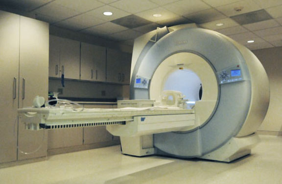
| MRI Machine |
Three-dimensional(3D)
imaging is important role in modern diagnostic radiology. Magnetic Resonance
Imaging (MRI) is one of the modern diagnostic radiology equipment that can
create images of the human body. No radiation used and it is non-invasive
because its only based on magnetic fields of the hydrogen atom in the body. MRI
can also produce computer-generated images of the body tissues and organs.
Basically, the images are two-dimensional(2D), where MRI produce images that
are presented in slices from top to bottom but these 2D images cannot be
produce to another plane without losing it significant image resolution. If the
information needed is in other planes, the acquisitions need to be repeated at
the planes needed. Since 2D imaging is not quite efficient, there is 3D MRI for
medical used for nowadays. The 3D data only acquired one plane and the images
for other planes also produced with the same 3D data set. In Addition, 3D
imaging also allows the presentation of anatomic detail so this make it easier
for the radiologist to manipulate images by removing or making them transparent
of unwanted overlying structures or emphasizing areas of interest. These
techniques make the radiologists and surgeons to understand anatomic
relationships better. This 3D imaging is more productive for planning of
surgical procedures such as preoperative evaluation of vascular and biliary
ductal variations in living-related liver donors, assessment of vascular
patency in patients undergoing surgical resection, preoperative liver and
tumour volume measurements and virtual hepatectomy.


No comments